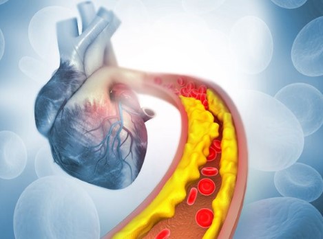
Atherosclerosis and the structural and functional state of the vessels of the carotid and vertebrobasillary basins
Post updated: July 19
Every year statistics show an increase in morbidity and mortality from cardiovascular diseases, primarily such as ischemic stroke and myocardial infarction.
It should be noted that the incidence and mortality from stroke in Ukraine is significantly higher than in Europe and the USA. Among all disorders of cerebral circulation (NMC), ischemic strokes are diagnosed in 75-80% of cases [1, 2, 19]. The most common cause of thromboocclusive lesions of the vascular system of the brain is atherothrombosis – a generalized and progressive process that depends on the evolution of atherosclerotic changes in the vessels.
According to the modern concept of atherosclerosis and atherothrombosis, the clinical manifestation of most cardiovascular catastrophes is directly related to the moment of violation of the integrity of the atherosclerotic plaque [3, 4]. A fundamentally important link in the pathogenesis of atherosclerosis and its complications (atherostenosis, atherothrombosis, atheroembolism, atheroocclusion, plaque hemorrhage followed by thromboembolism) is endothelial dysfunction. The endothelium (the inner lining of the vessels) consists of approximately 1.6×10¹³ cells, with a total weight of about 1 kg and a total area of ≈ 900 m2. Endotheliocytes have a pronounced metabolic activity and perform various functions. That is why the endothelium, in fact, can be considered as the largest endocrine gland.
The stages of the evolution of atherosclerotic plaque are associated with endothelial dysfunction. Endothelial dysfunction, according to the most modern hypothesis, develops due to chronic damage to it, which leads to platelet adhesion to the subendothelial layer and their aggregation, the release of growth factors that promote the migration of smooth muscle cells from media to intima with the formation of fibrous plaques. An important role in the mechanisms of atherogenesis is also played by the hemodynamic factor, manifested by the damaging local effect of the blood flow on the vessel wall, on its endothelium in places of physiological bends and bifurcations.
The widespread introduction of the latest methods of neuroimaging, including ultrasound, has allowed to increase the degree of detection of the atherosclerotic process in the vessels of the brain, as well as the percentage of their asymptomatic lesion.
In connection with modern pathogenetic ideas about the mechanisms of development of ischemic stroke, early diagnosis of this disease becomes even more important. The question of the informativeness of noninvasive ultrasound methods of investigation used to study the condition of cerebral arteries that are involved in blood supply to the brain is becoming relevant at the present stage [13, 17].
Objective: to study the structural and functional state of the vessels of the carotid and vertebrobasillary basins in elderly patients with cerebral atherosclerosis (CA) stages 1-3, including depending on the hemispheric localization of the ischemic focus.
Materials and methods.
A comprehensive clinical and instrumental study involved 229 patients with grade 2-3 CA. The diagnosis of "Cerebral atherosclerosis" was established in accordance with the classification of atherosclerosis of the World Health Organization from 2015 and was confirmed by laboratory and instrumental studies (ultrasound Dopplerography of cerebral arteries, magnetic resonance imaging (MRI) of the brain).
Study design: simple prospective non-randomized, with sequential inclusion of patients.
It was conducted on the basis of the Department of vascular Pathology of the brain of the State Institution "D.F. Chebotarev Institute of Gerontology of NAMS of Ukraine", Kiev.
All patients underwent conventional clinical, laboratory and instrumental examination (ultrasound Dopplerography of the vessels of the head and neck – examination of the cerebral blood flow of the extra- and intracranial sections of the main arteries of the head and neck on the Aplio XG (Toshiba) device, MRI of the brain).
See the full text of the article below.


